User:Raghavendra029/sandbox
HOMOCYSTINURIA[edit]
Introduction[edit]

Homocystinuria is a disorder of Methionine metabolism, leading to an abnormal accumulation of homocysteine and its metabolites (homocystine, homocysteine-cysteine complex, and others) in blood and urine. Normally, these metabolites are not found in appreciable quantities in blood or urine.
Homocystinuria is an autosomal recessively inherited defect in the transsulfuration pathway (homocystinuria I) or methylation pathway (homocystinuria II and III)
Pathophysiology[edit]
Homocysteine metabolism:
The accumulation of homocysteine and its metabolites is caused by disruption of any of the 3 interrelated pathways of methionine metabolism—deficiency in the cystathionine B-synthase (CBS) enzyme, defective methylcobalamin synthesis, or abnormality in methylene tetrahydrofolate reductase (MTHFR). Clinical syndromes resulting from each of these metabolic abnormalities have been termed homocystinuria I, II, and III. Three different cofactors/vitamins—pyridoxal 5-phosphate, methylcobalamin, and folate—are necessary for the 3 different metabolic paths. The pathway, starting at methionine, progressing through homocysteine, and onward to cysteine, is termed the transsulfuration pathway. Conversion of homocysteine back to methionine, catalyzed by MTHFR and methylcobalamin, is termed the remethylation pathway. A minor amount of remethylation takes place via an alternative route using betaine as the methyl donor. [2]
Epidemiology[edit]
The incidence of homocystinuria in the United States is approximately 1 per 100,000. Internationally, the reported incidence of homocystinuria varies between 1 in 50,000 and 1 in 200,000. Early diagnosis and intervention have helped in preventing some of the complications of homocystinuria, including ectopia lentis, mental retardation, and thromboembolic events. A mortality rate of 18% by age 30 has been reported by Mudd et al from a worldwide series of 629 patients with CBS enzyme deficiency. [4] Death is predominantly due to cerebrovascular or cardiovascular causes. Children with CBS deficiency (homocystinuria I) may be normal at birth. Data from Mudd et al suggest that starting at around age 20 years, these patients have an increasing likelihood of suffering a thromboembolic event. Patients with either defective methylcobalamin synthesis or defective tetrahydrofolate metabolism may present in early infancy.
Clinical Presentation[edit]
Homocystinuria is associated with the following physical findings:
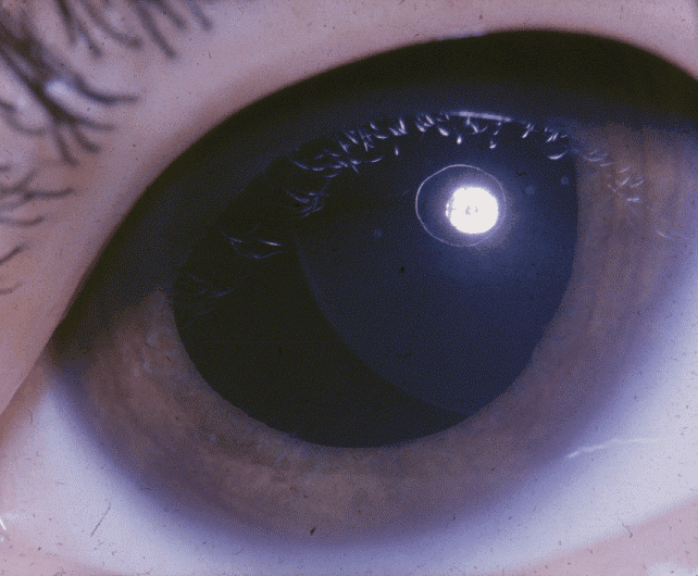
- Downward dislocation of lens (ectopia lentis)
Ectopia Lentis
- Marfanoid habitus
- Pes excavatum, pes carinatum, and genu valgum
- Mental retardation
- Signs and symptoms of strokes in any vascular distribution: hemiplegia, aphasia, ataxia, and pseudobulbar palsy are among the most common findings.
Patients with classic homocystinuria may first be recognized because of downward dislocation of the lens (ectopia lentis) [5] , marfanoid habitus, mental retardation [5] , and/or seizures. Patients with defective methylcobalamin synthesis may have all of these features, along with symptoms of methylmalonic acidemia (see Metabolic Disease and Stroke - Methylmalonic Acidemia). Acute stroke symptoms may occur in these patients. Traditional risk factors—hypertension, smoking, and diabetes—may or may not be present. The oral health of 14 patients with homozygote cystathionine beta synthase-deficient homocystinuria was evaluated in a Swedish study and found to be compromised in a majority of cases. The authors suggested that methionine restriction (low-protein diet) in the treatment of homocystinuria may result in a diet high in sugars. They therefore noted the need for regular dental checkups and preventive oral care for individuals suffering from homocystinuria. In addition, the authors noted that all patients had short dental roots, particularly of the central maxillary incisors. [6] In a Spanish cross-sectional survey sent to 35 hospitals, 75 patients were identified with homocystinuria: 41 with transsulfuration defects (1 death), 27 with remethylation defects (6 deaths), and 7 without a syndromic diagnosis. In 18 cases, more than one sibling was affected. Patients with remethylation defects had the most severe clinical manifestations. There was a high percentage of cognitive impairment, followed by lens diseases. Neurologic disorders were present in almost half of the patients, and there was increased vascular involvement in CBS-deficient adults.
Diagnosis[edit]
Several studies have pointed out that early diagnosis and institution of treatment and dietary restriction is likely to slow the progression of disease in homocystinuria as well as to reverse some of the features. If family history and sibling history suggest homocystinuria, screening tests should be advised. Early signs of myopia and lens abnormalities cannot be ignored. [8] Bony abnormalities and body habitus can be confused with Marfan syndrome; however, Marfan syndrome follows an autosomal dominant inheritance pattern, while homocystinuria follows a recessive pattern. The differential diagnosis includes the following:
- Blood Dyscrasias and Stroke
- Metabolic Disease & Stroke: Fabry Disease
- Metabolic Disease & Stroke: Methylmalonic Acidemia
- Metabolic Disease & Stroke: Propionic Acidemia
Other problems to be considered include carotid disease and stroke. Acute stroke diagnosis and treatment requires that certain laboratory studies such as complete blood count, chemistries, prothrombin/activated partial thromboplastin times (PT/aPTT), brain imaging, echocardiography, and vascular studies be done to exclude the usual causes, some of which may be treatable or preventable.
Brain Imaging:
If homocystinuria is suspected on the basis of history, physical examination, and family history, the patient may be transferred to a tertiary care center, where expertise in a variety of relevant fields is more likely to be available.
Laboratory studies for homocystinuria[edit]
If patients present with systemic signs and symptoms, screening tests followed by confirmatory tests may be done. The urine screening test for sulfur-containing amino acids, called the cyanide nitroprusside test, can be undertaken; however, high rates of false-negative as well as false-positive results are reported. A neonatal screening test, called the Guthrie test, detects high levels of methionine in heel-stick blood. This test is performed routinely in several states for detection of phenylalanine, leucine, and methionine. Because of high false-negative results in homocystinuric patients, a recent report suggested lowering the threshold of methionine to qualify as abnormal. Quantitative tests for homocystine in urine and blood are available commercially. The blood specimen needs to be handled in a specific manner described in the homocysteinemia testing section. Measurement of CBS activity in cultured fibroblasts provides definitive support for the diagnosis. Testing of amniotic cells and chorionic villi is also available.
Treatment[edit]
Pyridoxine, at a dose of 100-500 mg/d, is the drug of choice. Patients may be divided into pyridoxine-sensitive and pyridoxine-insensitive groups. In the first group, pyridoxine, folic acid, and vitamin B-12 are prescribed. These 3 vitamins, in combination, reduce the homocysteine levels as well as provide clinical benefit. Secondary stroke prevention rests on risk factor reduction. Aspirin, clopidogrel, and aspirin-dipyridamole have been suggested for secondary stroke prophylaxis, but whether other antiplatelet agents or anticoagulation are equally or more effective is not known. Measuring homocystine levels can be used to monitor the effectiveness of treatment. If pyridoxine alone is not effective, folic acid and vitamin B-12 can be added to the regimen. If patients are pyridoxine insensitive, a low-methionine diet initiated at diagnosis, along with betaine supplementation, may help reduce homocysteine levels. [10]
Consultations[edit]
An experienced neurologist (adult or pediatric) should be consulted both for acute care of a patient with a stroke and for the diagnosis of uncommon causes of a stroke. Genetic counseling should be offered to the patient and the family on confirmation of homocystinuria. Dietary consultation may be required if a homocystinuric patient is found to be pyridoxine insensitive and requires dietary modification. Physical, occupational, and speech therapists may be consulted for patients with acute stroke.
Prognosis[edit]
Early diagnosis of homocystinuria along with prophylactic medical and dietary care is a key to better long-term prognosis; it can halt or even reverse some of the complications.
Complications[edit]
Patients with homocystinuria are prone to thromboembolic events in the perioperative and postoperative periods, even with minor surgeries. Preoperative levels of homocysteine should be reduced to a near normal level. During and after surgery, aggressive hydration and prophylaxis for deep vein thrombosis (DVT) are strongly recommended.
Society and Culture[edit]
One theory suggests that Akhenaten, a pharaoh of the eighteenth dynasty of Egypt, may have suffered from homocystinuria.[10]
References[edit]
- ^ Yoo JH, Chung CS, Kang SS. Relation of plasma homocyst(e)ine to cerebral infarction and cerebral atherosclerosis. Stroke. 1998 Dec. 29(12):2478-83. [Medline].
- ^ Richard E, Desviat LR, Ugarte M, Pérez B. Oxidative stress and apoptosis in homocystinuria patients with genetic remethylation defects. J Cell Biochem. 2013 Jan. 114(1):183-91. [Medline]
- ^ Rozen R. Molecular genetic aspects of hyperhomocysteinemia and its relation to folic acid. Clin Invest Med. 1996 Jun. 19(3):171-8. [Medline].
- ^ Mudd SH, Skovby F, Levy HL, Pettigrew KD, Wilcken B, Pyeritz RE, et al. The natural history of homocystinuria due to cystathionine beta-synthase deficiency. Am J Hum Genet. 1985 Jan. 37(1):1-31. [Medline]. [Full Text].
- ^ Kaur M, Kabra M, Das GP, Suri M, Verma IC. Clinical and biochemical studies in homocystinuria. Indian Pediatr. 1995 Oct. 32(10):1067-75. [Medline].

