User:Anuragk007

Science: Control and Coordination
A system of control and coordination is essential in living organisms so that the different body parts can function as a single unit to maintain Homeostasis as well as respond to various stimuli.(Human homeostasis refers to the body's ability to regulate physiologically its inner environment to ensure its stability in response to fluctuations in the outside environment and the weather. Homeo=same and stasis=standing still) To carry out a simple function such as picking up an object from the ground there has to be coordination of the eyes,hands, legs and the vertebral column. The eyes have to focus on the object, the hands have to pick it up and grasp it, the legs have to bend and so does the back bone (vertebral column). All these actions have to be coordinated in such a manner that they follow a particular sequence and the action is completed. A similar mechanism is also needed for internal functions of the body.Some of these movements are in fact the result of growth, as in plants. A seed germinates and grows and we can see that the seedling moves over the course of a few days, it pushes soil aside and comes out.
7.1 ANIMAL-NERVOUS SYSTEM
Nervous system performs the main task of co-ordination among the internal organs as well as between the animal and its environment. In vertebrates, this system can be divided into (a) central, (b) peripheral (c) sympathetic (d) parasympathetic.
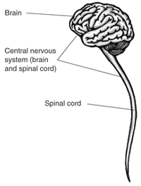
Central nervous system includes the brain and the spinal cord. The structural and functional unit of nervous system is the nerve cell called neuron. Neurons and neuralgia constitute the nervous tissue which is the prime tissue in the make up of the nervous system. Let us first study and understand the structural and functional unit of the nervous system.

Structure of Neuron[edit]
A nerve cell is an elongated cell which can be divided into 3 parts:
(i) Dendrite[edit]
It is a hair-like process which is hollow. It is connected to the cyton. The number of dendrites may be more than one. Dendrites may also be branched. Dendrites receive sensation or stimulus which may be physical, chemical, mechanical or electrical. It passes on the stimulus to the cyton.
(ii) Cyton[edit]
This part of the neuron has a central nucleus and surrounding cytoplasm. Around the nucleus there are granules called Nissl granules. Stimulus is changed in cyton to another form called impulse. From one side of cyton arises a cylindrical process filled with cytoplasm. This process is called axon.
(iii) Axon[edit]
It is the longest part of the neuron. It transmits the impulse from cyton to the tip of the axon called axon bulb. The axon is generally covered by a sheath of lipoprotein called myelin sheath. This sheath is formed by a type of cell called Schwann cell. At one point the axon is slightly depressed (a notch, an indentation). This is called the node of ranvier. When an impulse travels along the axon, an electromechanical change can be seen.
When the impulse is not travelling, the axon is said to be in the resting condition. In this condition, its outer part becomes positively charged. As the impulse begins travels, the positive charge comes inside and the outer surface becomes progressively negatively charged. After the impulse has travelled to the end point, a chemical compound, acetylcholine, is discharged from the axon bulb to the outside space, called synapse. Within milliseconds the charges in the axon are rearranged and the outer surface again becomes positively charged.
A neuron with one dendrite and one axon is called bipolar. A neuron, with two dendrites and one axon is called tripolar. The number of dendrites varies, but there is only one axon. In pentapolar neuron, there are four dendrites. The neuron in which axon is not covered by myelin sheath, is called non-medullated neuron.

Central Nervous System[edit]
In vertebrates, the central nervous system consists of brain, and the spinal cord.
Brain: -[edit]
It is the most important organ which is lodged in the brain box, called cranium. Brain is covered by membranes called meninges. Between the membranes and the brain and also inside the brain, there is a characteristics fluid, called cerebrospinal fluid. The brain can be divided into three main parts:
i. Fore-brain : -[edit]
This is the anterior part which includes (a) olfactory lobes, the centers of smell; (b) cerebral hemispheres, the seat of intelligence & voluntary action; (c) diencephalons, the centre of hunger, thirst, etc.
ii. Midbrain : -[edit]
This part includes optic lobes which are centre of vision.
iii. Hind-brain :-[edit]
This is the posterior part which includes the (a) cerebellum, the co-ordination centre of involuntary actions. Medulla oblongata is continued behind into the spinal cord. The brain is hollow. It has four longitudinal cavities called ventricles. In land vertebrates, 12 different cranial nerves are connected with the brain. Spinal cord It is a long cord which arises from medulla oblongata and runs all along the vertebral column. It pass through the neural canal which is a canal of vertebra.
In the transverse section of a spinal cord, a central canal can be seen. This canal remains filled with cerebrospinal fluid. Immediately surrounding the canal, there are clusters of cytons which form the grey matter. In the peripheral part, axons are concentrated and, therefore, this area is called white matter. From each side of spinal cord, there are two horns, the dorsal horn and the ventral horn. To the dorsal horn joins a nerve which picks up sensation from the organ. It is called sensory organs. From the ventral horn or root arises the motor nerve which takes the message from the spinal cord to organ concerned. These two nerves constitute the reflex action. This action is very quick, for example, movements of eyelids, sneezing, coughing, yawning, hiccupping, shivering etc. In man 31 pairs of spinal nerves can be seen; eight in the neck region, 12 in chest region, five in abdominal region, five in hip region, and one in coccyx region. Coccyx is the last bone of the vertebral column.
Autonomic nervous system[edit]
Nerves from the brain and the spinal cord connect the skeletal muscles and control their activities according to the direction and demand of the body. These nervous are, therefore related to the voluntary acts, i.e., acts according to the desire. But the internal organs are not under the control of our will. We can not rotate the stomach or accelerate the heart beat by our conscious effort. For the control of the activities of the internal organs there is another type of nervous system called autonomous nervous system. You have seen the case of reflex action that motor neuron arises from the spinal cord and pass along the ventral root uninterruptedly all the way to the skeletal muscle. The cyton of this neuron is located in the spinal cord. But in case of an autonomous nervous system, i.e., spinal cord. This neuron runs for some distance only. It terminates in the sympathetic ganglion where it passes through the massage to the second nerve or neuron through a synapse which carries the impulse to the muscle or gland.
The autonomous nervous system has two subdivisions: sympathetic and parasympathetic. The sympathetic nervous system originates from the thoracic (chest) and lumber (abdominal) areas of spinal cord. Parasympathetic nerve which arises from spinal cord runs for a considerable distance and it forms a synapse (the point of exchange of impulse from one nerve to another) near the target organ where it has to send the message. The sympathetic nerves which arise from the spinal cord form a synapse in regular chain of ganglia running parallel to the spinal cord.
Sympathetic and parasympathetic nervous systems function in just the opposite manner. For example, if a person become angry, it may be due to the discharge of a chemical to different organs by the sympathetic nerves leading to increased heartbeat, etc. the parasympathetic nerve may discharge a different chemical, and thereby slow down the heartbeat and bring the person to normal state.

7.1.1 WHAT HAPPENS IN REFLEX ACTION?
The Reflex ArcIf you stand on something sharp then you move your foot very quickly. You don't have to think about it - it feels like it happens even before you feel the pain. This is because the process is a reflex and doesn't involve the brain. Receptors and Effectors. The diagram shows someone stepping on a drawing pin. Receptors in the skin of the foot will register the fact that this has happened and will send a signal along a sensory neuron to the spinal cord. Inside the spinal cord an interneuron will transfer the nerve signal to another nerve cell. This cell is a motor neuron and it carries the signal to the muscles in the leg. The leg muscles will contract and, hopefully, the foot will be moved away from the source of pain.
Each of the nerve cells are separated by synapses. The Patellar Reflex (or Knee-Jerk)You may have seen doctors on the television (or maybe it's happened to you) where they test someone's reflexes by tapping just under the kneecap (patella) with a small rubber hammer. If this spot is hit just right then it will cause some special sensory cells (called a muscle spindle) to send signals off to the spinal cord. The signal passes through the interneuron and then via a motor neuron to the quadriceps muscle. This causes the leg to kick forward.
No matter how much you concentrate you cannot stop this from happening as it's a reflex action and doesn't involve the brain. did you know ...The Latin name for the common limpet is Patella vulgata. This is because the shell of the limpet does look a bit like a kneecap.
