User:Gorozco1/sandbox
It is generally understood that the process regulating wakefulness and sleep is driven by circadian rhythms influenced by light-dark cycles. Even though it has been shown that this innate biological clock increases the physiological propensity for sleep during the later hours of the day when the sun is absent, a focus on this concept alone would afford only one-half of the underlying mechanism. The regulation of the sleep-wake cycle is a process warranting an explanation beyond what is offered by a simple discussion of circadian rhythms, and, as the title of this article has already insinuated, can be explained by a treatment of a two-process model: process-S and process-C. Process-S is less commonly known and represents a homeostatic drive for sleep that progressively increases during wakefulness decreasing only during NREM sleep; furthermore, process-C, which is more commonly known as circadian rhythms, represents a 24-hour internal oscillation of endogenous, entrainable bodily rhythms.[1] These two processes work in a synergistic way to promote sleep by combining naturally occurring rhythms with a homeostatic pressure system facilitated by the neuromodulator adenosine.
Process-S[edit]
Process-S is a homeostatic drive, a kind of pressure system, that builds up during the wakeful hours in a person's day. This sleep pressure rises during wakefulness, drops exponentially during sleep, and continues to increase when sleep deprivation occurs.[2] Once a person falls asleep, it has been empirically measured by a technique using electroencephalography (EEG) that the level of sleep pressure correlates with increased presence of slow-wave activity in the first half of a sleep bout.[3] This theory is found to be consistent with contemporary hypnogram models, which show the first half of a sleep episode primarily dominated by stage-3 and stage-4 slow-wave sleep; furthermore; hypnograms of sleep deprived participants have been shown to have increased amounts of stage-3 and stage-4 sleep during the first half of a sleep episode meaning increased duration of wakefulness corresponds to increased duration of slow-wave sleep.[4]
Hypnogram[edit]

Electrical brain activity progresses throughout a sleep episode in ultradian cycles of rapid-eye movement sleep (REM) and non-rapid eye movement sleep (NREM).[5] As the electrical activity in the brain continues, the relationship between stage-3 and stage-4 slow-wave sleep and REM sleep exhibits a negative correlation. As Figure 1. on the right shows, stage-3 and stage-4 sleep dominate the first half of sleep while REM (marked in red) only accounts for a small amount. In the second half of a sleep episode, slow-wave sleep is non-existent and REM sleep dominates along with stage-1 and stage-2, both of which are not considered slow wave sleep. The peaks and trough of this graph represent an ultradian cycle that follows a specific pattern depending on the temporal stage of sleep: S1→S2→S3→S4→S3→S2→REM cycle for the first half of a sleep episode, and S1→S2→S1→REM for the second half.
How process-S is incorporated into this hypnogram is quite elegant when using sleep deprived individuals as an example. If a person is deprived of sleep and their homeostatic pressure is allowed to built up over the course of a day, they should feel a strong urge to sleep (makes sense right?). Once asleep, not only does the person experience a longer sleep episode, but they also experience more intense uninterrupted sleep.[6] This research by Borbely & Achermann (2003) has shown that a sleep deprived individual will spend increased amounts of slow-wave sleep early in the sleep episode to compensate for the sleep pressure built up during the day, which is electronically represented by the troughs and the peaks of the hypnogram during the first half of the sleep episode.
Adenosine[edit]

Adenosine is a neuromodulator found throughout the brain. The homeostatic pressure present in process-S has been directly correlated to the build up of adenosine, which means that the presence of a larger sleep pressure equals higher amounts of circulating adenosine. Concentrations of adenosine in the brain gradually increases over time as a result of ongoing metabolic activity in the brain.[7] The build up of this neuromodulator over the course of a day has the effect of decreasing wakefulness, and is only halted when metabolic activity is reduced (i.e. sleep). A study by Porkka-Heiskanen et al (2000) has shown that during NREM sleep metabolic activity decreases 30% facilitating the release of this built up homeostatic pressure and further implicating adenosine as a mediator of sleepiness following prolonged wakefulness. [8] Rat studies incorporating the use of adenosine further supports the theory of adenosine as a supporting facilitator of sleep. In these studies, rats were administered adenosine producing symptoms of sedation, sleep, and increases in SWS.[9]
Caffeine[edit]
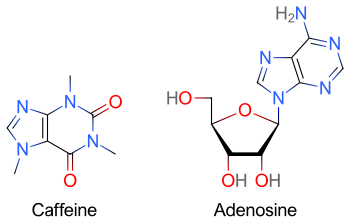
The molecule caffeine is an alkaloid that belongs to a group called the xanthines.[10] The psychoactive effect of this substance can be largely attributed to its antagonistic effect on adenosine receptors producing the stimulating state so often described by its consumers. Moderate doses of caffeine, ranging from 200-350 mg, produce physiological and psychological effects such as elevated blood pressure and heart rate, improved mood and concentration, and increased energy and wakefulness.[11] This range of intake is considered to be the therapeutic dose of caffeine due to its positive influence on concentration and energy and its stimulation of the sympathetic nervous system. The chemical structure of caffeine, as shown in Figure 2, has a somewhat similar shape to the molecule adenosine which allows it to dock in its receptors and produce its well known psychoactive effect. Upon docking with adenosine receptors, the caffeine molecule acts as an antagonist prohibiting the receptor to be stimulated by its own agonist.
Clinical trials further implicate caffeines effect on wakefulness. It has been well researched that Parkinson's disease (PD) is associated with sleep abnormalities such as excessive daytime sleepiness (EDS). EDS in PD individuals is defined as spontaneous dozing during the day that occurs in 50% of the PD population.[12] EDS is almost analogous to narcolepsy episodes in the sense that sufferers from this syndrome can fall asleep during life threatened motor operations such as driving a car. In a study by Knie et al. (2011), it has been shown that the onset of EDS is correlated with deterioration in the nigrostriatal system and extrastriatal neurons in the mid/hind brain.[13] . Cauli & Morelli (2005) have shown that when the effects of adenosine are blocked in these regions of the brain, an increased release of the neurotransmitter glutamate is allowed to exert its influence on the striatum promoting the activity of the neurons regulating motor function and behavior.[14] This means that adenosine protects dopaminergic neurons from apoptosis but in an indirect way (by being absent). Motor functioning facilitated by caffeine through the use of glutamate just proves the already known interconnectivity of the brain, and corroborates with the theory that adenosine is significantly related to the slowing of the homeostatic build-up known as process-S, which is shown by its effectiveness on treating EDS.
Process-C[edit]
Process-C, also known as circadian rhythms (circa = about, dies = day), is the body's internal clock and has been localized to the suprachiasmatic nucleus (SCN).[15] Process-C is independent of sleep and waking unlike process-S, and relies on external cues known as a zeitgeber.[16] To classify a biological rhythm as circadian, the following criteria must be met:
- The rhythm must have a 24-hour cycle
- The rhythm is said to be endogenous meaning even during free-running experiments the rhythms persist even if slightly extended.
- The rhythm can be entrained through the use of external cues such as light known as zeitgebers.
-For a more extensive description visit the circadian rhythms wikipedia page
Process-S x Process-C Interaction[edit]

Both circadian processes and homeostatic processes contribute equally to the two-process model of sleep regulation.[17] This model is represented by Figure 3, and at first glance the fluctuations between the two processes is the first thing warranting a discussion. The circadian wake drive represents process-C and its sine wave shape never changes, though it can be phase shifted earlier or later. The homeostatic sleep drive represents process-S and its fluctuations represents increases and decreases in adenosine due to metabolic activity in the brain. The way these two processes synergistically affect sleep behavior is simply a matter of mathematical difference at any given time point, but for clarity reasons only two times will be discussed: 7 a.m. and 11 p.m.. At 7 a.m. circadian rhythms encourage wakefulness usually by means of light stimuli (sunrise). As the day progresses and the day becomes darker, one begins to feel sleepy as melatonin secreted by the pineal gland is released.[18] Along with this process, homeostatic pressure is built up throughout the day until the two processes collectively create a sense of sleep necessity. At any given time between these two processes, the measure of sleepiness can be determined from the distance between the circadian sine wave and the homeostatic sleep curve. This means that at 11 p.m. the distance between the two graphs is the greatest so the urge to sleep should also be the greatest. Once sleep is initiated and process-S is allowed to decrease in pressure, the distance between the two graphs is reduced. Sleep pressure is continually decreased until the point of contact with process-C which signals the body to move to a state of wakefulness consequently initiating the increasing trend of process-S all over again. One could imagine what the graph would look like if sleep deprivation occurred. If a person were to stay up all night and not receive the depressurization effect of process-S by sleep, the distance between the two graphs the following day would be so great at 11 p.m. that an extremely strong push for sleep would be experienced.
- ^ Van Dongen, Hans P. A.; Dinges, David F. (26). "Investigating the interaction between the homeostatic and circadian processes of sleep–wake regulation for the prediction of waking neurobehavioural performance". Journal of Sleep Research. 12 (3): 181–187. doi:10.1046/j.1365-2869.2003.00357.x. PMID 12941057.
{{cite journal}}: Check date values in:|date=and|year=/|date=mismatch (help); Unknown parameter|month=ignored (help) - ^ Taillard, Jacques; Philip, Pierre; Coste, Olivier; Sagaspe, Patricia; Bioulac, Bernard (29). "The circadian and homeostatic modulation of sleep pressure during wakefulness differs between morning and evening chronotypes". Journal of Sleep Research. 12 (4): 275–282. doi:10.1046/j.0962-1105.2003.00369.x. PMID 14633238.
{{cite journal}}: Check date values in:|date=and|year=/|date=mismatch (help); Unknown parameter|month=ignored (help) - ^ Crowley, Stephanie J.; Acebo, Christine; Carskadon, Mary A. (2007). "Sleep, circadian rhythms, and delayed phase in adolescence". Sleep Medicine. 8 (6): 602–612. doi:10.1016/j.sleep.2006.12.002. PMID 17383934.
{{cite journal}}: CS1 maint: date and year (link) - ^ Durmer, Jeffrey S.; Dinges, David F. (2005). "Neurocognitive Consequences of Sleep Deprivation". Seminars in Neurology. 25 (1): 117–129. doi:10.1055/s-2005-867080. PMID 15798944.
{{cite journal}}: CS1 maint: date and year (link) - ^ Achermann, Peter; Borbély, A. A. (1). "Mathematical Models of Sleep Regulation". Frontiers in Bioscience. 8 (6): s683-693. doi:10.2741/1064. PMID 12700054.
{{cite journal}}: Check date values in:|date=and|year=/|date=mismatch (help); Unknown parameter|month=ignored (help) - ^ Achermann, Peter; Borbély, A. A. (1). "Mathematical Models of Sleep Regulation". Frontiers in Bioscience. 8 (6): s683-693. doi:10.2741/1064. PMID 12700054.
{{cite journal}}: Check date values in:|date=and|year=/|date=mismatch (help); Unknown parameter|month=ignored (help) - ^ Porkka-Heiskanen, Tarja; Strecker, Robert E.; Thakkar, Mahesh; Bjørkum, Alvhild A.; Greene, Robert W.; McCarley, Robert W. (23). "Adenosine: A Mediator of the Sleep-Inducing Effects of Prolonged Wakefulness". Science. 276 (5316): 1265–1268. doi:10.1126/science.276.5316.1265. PMC 3599777. PMID 9157887.
{{cite journal}}: Check date values in:|date=and|year=/|date=mismatch (help); Unknown parameter|month=ignored (help) - ^ Porkka-Heiskanen, T.; Strecker, R.E; McCarley, R.W (16). "Brain site-specificity of extracellular adenosine concentration changes during sleep deprivation and spontaneous sleep: an in vivo microdialysis study". Neuroscience. 99 (3): 507–517. doi:10.1016/S0306-4522(00)00220-7. PMID 11029542.
{{cite journal}}: Check date values in:|date=and|year=/|date=mismatch (help); Unknown parameter|month=ignored (help) - ^ Ticho, Simon R.; Radulovacki, M. (1991). "Role of Adenosine in Sleep and Temperature Regulation in the Preoptic Area of Rats". Pharmocology Biochemistry & Behavior. 40 (1): 33–40. doi:10.1016/0091-3057(91)90317-U. PMID 1780343.
{{cite journal}}: CS1 maint: date and year (link) - ^ Kalow, Werner; Tang, Bing-Kou (1991). "Use of caffeine metabolite ratios to explore CYP1A2 and xanthine oxidase activities". Clinical Pharmacology and Therapeutics. 50 (5/1): 508–519. doi:10.1038/clpt.1991.176. PMID 1934864.
{{cite journal}}: CS1 maint: date and year (link) - ^ Temple, Jennifer L.; Dewey, Amber M.; Briatico, Laura N. (December 2010). "Effects of acute caffeine administration on adolescents". Experimental and Clinical Psychopharmacology. 18 (6): 510–520. doi:10.1037/a0021651. PMID 21186925.
{{cite journal}}: CS1 maint: date and year (link) - ^ Poewe, Werner; Högl, Birgit (August 2000). "Parkinson's disease and sleep". Current Opinion in Neurology. 13 (4): 423–426. doi:10.1097/00019052-200008000-00009. PMID 10970059.
{{cite journal}}: CS1 maint: date and year (link) - ^ Knie, Bettina; Mitra, M. Tanya; Logishetty, Kartik; Chaudhuri, K. Ray (1). "Excessive daytime sleepiness in patients with Parkinson's disease". CNS Drugs. 25 (3): 203–212. doi:10.2165/11539720-000000000-00000. PMID 21323392.
{{cite journal}}: Check date values in:|date=and|year=/|date=mismatch (help); Unknown parameter|month=ignored (help) - ^ Cauli, O.; Morelli, M. (15). "Caffeine and the dopaminergic system". Behavioural Pharmacology. 16 (2): 63–77. doi:10.1097/00008877-200503000-00001. PMID 15767841.
{{cite journal}}: Check date values in:|date=and|year=/|date=mismatch (help); Unknown parameter|month=ignored (help) - ^ Mistlberger, Ralph E. (7). "Circadian regulation of sleep in mammals: Role of the suprachiasmatic nucleus". Brain Research Reviews. 49 (3): 429–454. doi:10.1016/j.brainresrev.2005.01.005. PMID 16269313.
{{cite journal}}: Check date values in:|date=and|year=/|date=mismatch (help); Unknown parameter|month=ignored (help) - ^ Borb, Alexander A.; Achermann, Peter (December 1999). "Sleep Homeostasis and Models of Sleep Regulation". Journal of Biological Rhythms. 14 (6): 559–570. doi:10.1177/074873099129000894. PMID 10643753.
{{cite journal}}: CS1 maint: date and year (link) - ^ Borbely, Alexander A. (1982). "A two process model of sleep regulation". Human Neurobiology. 1: 166–178.
- ^ Crowley, Stephanie J.; Acebo, Christine; Carskadon, Mary A. (2007). "Sleep, circadian rhythms, and delayed phase in adolescence". Sleep Medicine. 8 (6): 602–612. doi:10.1016/j.sleep.2006.12.002. PMID 17383934.
{{cite journal}}: CS1 maint: date and year (link)
