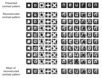
File:Visual stimulus reconstruction using fMRI.png
Visual_stimulus_reconstruction_using_fMRI.png (360 × 276 pixels, file size: 94 KB, MIME type: image/png)
 | This work is copyrighted (or assumed to be copyrighted) and unlicensed. It does not fall into one of the blanket acceptable non-free content categories listed at Wikipedia:Non-free content § Images or Wikipedia:Non-free content § Audio clips, and it is not covered by a more specific non-free content license listed at Category:Wikipedia non-free file copyright templates. However, it is believed that the use of this work:
qualifies as fair use under United States copyright law. Any other uses of this image, on Wikipedia or elsewhere, may be copyright infringement. See Wikipedia:Non-free content and Wikipedia:Copyrights. |
| Description |
Visual stimulus reconstruction using fMRI, by the ATR Computational Neuroscience Laboratories in Kyoto, Japan |
|---|---|
| Source |
Part of the article "Visual Image Reconstruction from Human Brain Activity using a Combination of Multiscale Local Image Decoders", published in the journal Neuron on 10 December 2008. direct link to image |
| Article | |
| Portion used |
1/4 of the original image |
| Low resolution? |
yes |
| Purpose of use |
to illustrate the article |
| Replaceable? |
no free alternative available |
| Other information |
Authors of the article: Yoichi Miyawaki, Hajime Uchida, Okito Yamashita, Masa-aki Sato, Yusuke Morito, Hiroki C. Tanabe, Norihiro Sadato, and Yukiyasu Kamitani. |
| Fair useFair use of copyrighted material in the context of Brain–computer interface//en.wikipedia.org/wiki/File:Visual_stimulus_reconstruction_using_fMRI.pngtrue | |
File history
Click on a date/time to view the file as it appeared at that time.
| Date/Time | Thumbnail | Dimensions | User | Comment | |
|---|---|---|---|---|---|
| current | 06:42, 2 December 2017 |  | 360 × 276 (94 KB) | Theo's Little Bot (talk | contribs) | Reduce size of non-free image (BOT - disable) |
| 14:41, 15 March 2009 | No thumbnail | 430 × 330 (109 KB) | Waldyrious (talk | contribs) | {{Non-free use rationale | Description = Visual stimulus reconstruction using fMRI, by the ATR Computational Neuroscience Laboratories in Kyoto, Japan | Source = Part of the article "Visual Image Reconstruction from Human Bra |
You cannot overwrite this file.

