
File:Staphylococcus aureus appearance on agar plates.jpg

Original file (2,370 × 1,368 pixels, file size: 453 KB, MIME type: image/jpeg)
| This is a file from the Wikimedia Commons. Information from its description page there is shown below. Commons is a freely licensed media file repository. You can help. |
Summary
| Description |
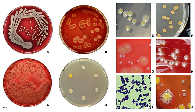
English: A Columbia blood agar plate with 5% sheep blood showing S. aureus subsp. aureus after 24 h of incubation. The normal phenotype is grayish colonies with a yellowish tint surrounded by a hemolysis zone.
B S. aureus with a wide zone of beta (complete) haemolysis on blood agar. The clearing of the erythrocytes in the agar is demonstrated by text “beta-hemolysis”underneath the plate being easily readable on spots where colonies were removed. C Protein toxins forming pores in biological membranes occur frequently in Gram-positive and Gram-negative bacteria. Because erythrocytes have often been used to test the activity of these toxins, some of them are also called hemolysins. Some strains of S. aureus can produce more than one hemolysin. Here you can see effect of various hemolysins on sheep erythrocytes. Colonies are surrounded by the zone of complete hemolysis continuing in three zones of incomplete lysis of sheep erythrocytes. D Pigment production in six strains of S. aureus on tryptic soy agar (TSA). 20 μl of bacterial suspension inoculated on the agar plate. S. aureus grows readily on many types of media producing pigments that vary from white to deep yellow. Many colonies develop pigment only upon prolonged incubation. E Colonies of two strains of S. aureus on tryptic soy agar. These two strains differ in colony size and pigment production. Both colony types are low-convex, smooth, and opaque. F Colonies of S. aureus on tryptic soy agar seen with transmitted light. Note that the distinctive golden pigment is most intense in the centre of the colony. Yellow pigment of S. aureus, staphyloxanthin, behaves as a virulence factor that helps the organism evade the immune system of the host. G Comparison of S. aureus and S. epidermidis colonies growing on Columbia sheep blood agar. S. aureus colonies are yellow pigmented and surrounded by a zone of beta-hemolysis. S. epidermidis colonies are without yellow pigmentation and non-hemolytic. H Many clinical isolates of S. aureus grow on cultivation media without conspicuous yellow pigmentation. Colonies seem to be whitish, but, unlike most other coagulase-negative staphylococci growing on sheep blood agar, they are surrounded by a zone of more or less profound beta-hemolysis. I S. aureus is a Gram positive bacterium. In the picture you can see Gram-positive spherical cells about 1 μm in diameter, non-sporing, occuring as single cocci, in pairs, as tetrads, or as short chains, which characteristically divide in more than one plane forming irregular clusters like a bunch of grapes. Gram stains should be performed on young cultures, because very old cells may lose their ability to retain crystal violet and may appear gram-variable or gram-negative. J S. aureus is a catalase-positive bacterium (yellow pigmented colonies) which means it can degrade hydrogen peroxide into water and oxygen, creating bubbles in 3% hydrogen peroxide solution. For example enterococci and streptococci are catalase negative. Non-pigmented grayish E. faecalis colony with the absence of bubble formation illustrates a negative catalase test performed directly on the agar plate. |
| Date | |
| Source | Own work |
| Author | HansN. |
Created for Wikipedia.org by HansN.
Licensing
- You are free:
- to share – to copy, distribute and transmit the work
- to remix – to adapt the work
- Under the following conditions:
- attribution – You must give appropriate credit, provide a link to the license, and indicate if changes were made. You may do so in any reasonable manner, but not in any way that suggests the licensor endorses you or your use.
File history
Click on a date/time to view the file as it appeared at that time.
| Date/Time | Thumbnail | Dimensions | User | Comment | |
|---|---|---|---|---|---|
| current | 16:19, 4 June 2019 | | 2,370 × 1,368 (453 KB) | New HanseN. | User created page with UploadWizard |
File usage
Global file usage
The following other wikis use this file:
- Usage on bn.wikipedia.org
- Usage on bs.wikipedia.org
- Usage on el.wikipedia.org
- Usage on hy.wikipedia.org
- Usage on ml.wikipedia.org
- Usage on sr.wikipedia.org