
File:RLS 12blauLeg.png
Size of this preview: 397 × 599 pixels. Other resolutions: 159 × 240 pixels | 318 × 480 pixels | 883 × 1,332 pixels.
Original file (883 × 1,332 pixels, file size: 1.56 MB, MIME type: image/png)
| This is a file from the Wikimedia Commons. Information from its description page there is shown below. Commons is a freely licensed media file repository. You can help. |
Summary
| Description |
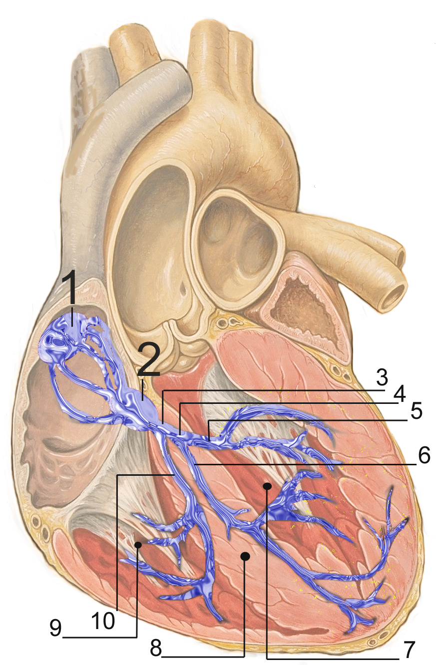
English: Electrical conduction system of the heart:
Français : Répartition du tissus nodal dans le coeur:
Русский: Проводящая система сердца:
Español: Sistema de conducción del corazón:
Deutsch: Erregungsleitungssystem:
Polski: Układ bodźcotwórczo-przewodzący:
|
| Date | |
| Source | self made, based upon Image:Heart anterior view coronal section.jpg by Patrick J. Lynch (Patrick J. Lynch; illustrator; C. Carl Jaffe; MD; cardiologist Yale University Center for Advanced Instructional Media ) |
| Author | J. Heuser |
| Permission (Reusing this file) |
Creative Commons Attribution 2.5 License 2007 |
preserve our creative credits: Patrick J. Lynch, medical illustrator; C. Carl Jaffe, MD, cardiologist. https://creativecommons.org/licenses/by/2.5/
Licensing
This file is licensed under the Creative Commons Attribution 2.5 Generic license.
- You are free:
- to share – to copy, distribute and transmit the work
- to remix – to adapt the work
- Under the following conditions:
- attribution – You must give appropriate credit, provide a link to the license, and indicate if changes were made. You may do so in any reasonable manner, but not in any way that suggests the licensor endorses you or your use.
File history
Click on a date/time to view the file as it appeared at that time.
| Date/Time | Thumbnail | Dimensions | User | Comment | |
|---|---|---|---|---|---|
| current | 17:51, 2 March 2007 | | 883 × 1,332 (1.56 MB) | JHeuser | {{Information |Description = Heart; conduction system |Source = self made, based upon Image:Heart anterior view coronal section.jpg by Patrick J. Lynch (Patrick J. Lynch; illustrator; C. Carl Jaffe; MD; cardiologist Yale University Center for Advanc |
File usage
The following pages on the English Wikipedia use this file (pages on other projects are not listed):
Global file usage
The following other wikis use this file:
- Usage on ar.wikipedia.org
- Usage on az.wikipedia.org
- Usage on bg.wikipedia.org
- Usage on bs.wikipedia.org
- Usage on ca.wikipedia.org
- Usage on cs.wikipedia.org
- Usage on de.wikipedia.org
- Usage on el.wikipedia.org
- Usage on es.wikipedia.org
- Usage on fa.wikipedia.org
- Usage on fr.wikipedia.org
- Usage on he.wikipedia.org
- Usage on id.wikipedia.org
- Usage on ko.wikipedia.org
- Usage on lt.wikipedia.org
- Usage on lv.wikipedia.org
- Usage on ml.wikipedia.org
- Usage on or.wikipedia.org
- Usage on pl.wikipedia.org
- Usage on pt.wikipedia.org
- Usage on ru.wikipedia.org
- Usage on sh.wikipedia.org
- Usage on sk.wikipedia.org
- Usage on sr.wikipedia.org
- Usage on ta.wikipedia.org
- Usage on th.wikipedia.org
- Usage on uk.wikipedia.org
- Usage on uz.wikipedia.org
- Usage on www.wikidata.org