
File:Mixed Tumor of the Salivary Gland.jpg
Mixed_Tumor_of_the_Salivary_Gland.jpg (550 × 361 pixels, file size: 33 KB, MIME type: image/jpeg)
| This is a file from the Wikimedia Commons. Information from its description page there is shown below. Commons is a freely licensed media file repository. You can help. |
Summary
| Description |
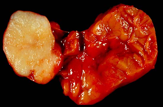
Mixed Tumor of the Salivary Gland This benign tumor of the submandibular gland, also known as pleomorphic adenoma, presented as a painless neck mass in a 40-year-old man. At the left of the image is the white tumor with its characteristic cartilaginous cut surface. To the right is the normally lobated submandibular salivary gland. Unlike most of my gross photos, this one was shot in the fresh state. This does show best what tissue looks like to the operating surgeon, but the soft, collapse-prone tissue, with its blood staining and distracting highlights, doesn't show anatomic details as well as do photos of formalin-fixed specimens. This pic was shot with a Minolta X-370 with a 100-mm Rokkor bellows lens, on Kodak Elite daylight ISO 100 film, using a blue compensator to correct for tungsten illumination. Photograph by Ed Uthman, MD. Public domain. Posted 13 May 00 |
| Source | http://web2.airmail.net/uthman/specimens/index.html |
| Author | |
| Permission (Reusing this file) |
PD |
Licensing
| This work has been released into the public domain by its author, Ed Uthman. This applies worldwide. In some countries this may not be legally possible; if so: Ed Uthman grants anyone the right to use this work for any purpose, without any conditions, unless such conditions are required by law.
|
File history
Click on a date/time to view the file as it appeared at that time.
| Date/Time | Thumbnail | Dimensions | User | Comment | |
|---|---|---|---|---|---|
| current | 09:51, 5 June 2006 | | 550 × 361 (33 KB) | Patho | {{Information| |Description=Mixed Tumor of the Salivary Gland This benign tumor of the submandibular gland, also known as pleomorphic adenoma, presented as a painless neck mass in a 40-year-old man. At the left of the image is the white tumor with its ch |
File usage
Global file usage
The following other wikis use this file:
- Usage on ca.wikipedia.org
- Usage on de.wikipedia.org
- Usage on de.wikibooks.org
- Usage on pl.wikipedia.org
- Usage on pt.wikipedia.org
- Usage on sr.wikipedia.org