
File:Diagram human cell nucleus numbered version.svg

Size of this PNG preview of this SVG file: 462 × 378 pixels. Other resolutions: 293 × 240 pixels | 587 × 480 pixels | 939 × 768 pixels | 1,252 × 1,024 pixels | 2,503 × 2,048 pixels.
Original file (SVG file, nominally 462 × 378 pixels, file size: 88 KB)
| This is a file from the Wikimedia Commons. Information from its description page there is shown below. Commons is a freely licensed media file repository. You can help. |
| Description |
Afrikaans: diagram van 'n selkern:
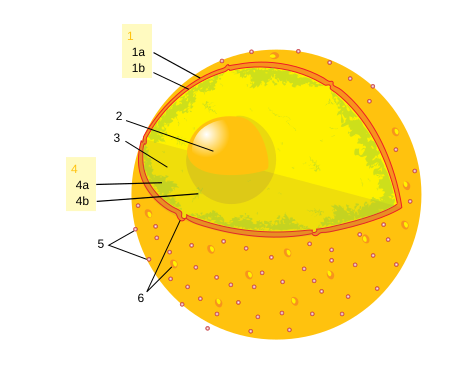
English: diagram of an cell nucleus:
Français : schéma d'un noyau cellulaire:
Polski: schemat jądra komórkowego.
Português: diagrama de um núcleo celular.
|
||
| Date | |||
| Source | File:Diagram human cell nucleus.svg | ||
| Author |
original version by Mariana Ruiz. numbered version by Felipe Fontoura. |
||
| Permission (Reusing this file) |
|
||
| Other versions |
[]
|
||
| SVG development |


.svg)

File history
Click on a date/time to view the file as it appeared at that time.
| Date/Time | Thumbnail | Dimensions | User | Comment | |
|---|---|---|---|---|---|
| current | 13:53, 26 May 2023 | | 462 × 378 (88 KB) | Puck04 | fixed SVG code (was "W3C-invalid"), cleanup, text elemets instead of path text |
| 19:34, 22 May 2007 |  | 462 × 378 (162 KB) | LipeFontoura | {{Information |Description = {{en|diagram of an cell nucleus: :'''1.''' Nuclear envelope. :'''1.a.''' Outer membrane. :'''1.b.''' Inner membrane. :'''2.''' Nucleolus. :'''3.''' Nuceleoplasm. :'''4.''' Chromatin. :'''4.a.''' Heterochro | |
| 19:06, 22 May 2007 |  | 462 × 378 (148 KB) | LipeFontoura | {{Information |Description = {{en|diagram of an en:cell nucleus.}} {{pt|diagrama de um pt:núcleo celular.}} |Source = Image:Diagram_human_cell_nucleus.svg |Date = 2007-05-22 |Author = original version by [[User:Ladyo |
File usage
The following pages on the English Wikipedia use this file (pages on other projects are not listed):
Global file usage
The following other wikis use this file:
- Usage on af.wikipedia.org
- Usage on be-tarask.wikipedia.org
- Usage on de.wikipedia.org
- Usage on fa.wikipedia.org
- Usage on fi.wikipedia.org
- Usage on he.wikipedia.org
- Usage on he.wiktionary.org
- Usage on hi.wikipedia.org
- Usage on id.wikipedia.org
- Usage on ja.wikipedia.org
- Usage on lv.wikipedia.org
- Usage on pl.wikipedia.org
- Usage on pnb.wikipedia.org
- Usage on rue.wikipedia.org
- Usage on se.wikipedia.org
- Usage on si.wikipedia.org
- Usage on te.wikipedia.org
- Usage on th.wikipedia.org
- Usage on uk.wikipedia.org
- Usage on ur.wikipedia.org
- Usage on vi.wikipedia.org
- Usage on zh.wikipedia.org