
File:Axoneme cross-section.svg

Size of this PNG preview of this SVG file: 600 × 600 pixels. Other resolutions: 240 × 240 pixels | 480 × 480 pixels | 768 × 768 pixels | 1,024 × 1,024 pixels | 2,048 × 2,048 pixels | 656 × 656 pixels.
Original file (SVG file, nominally 656 × 656 pixels, file size: 162 KB)
| This is a file from the Wikimedia Commons. Information from its description page there is shown below. Commons is a freely licensed media file repository. You can help. |
Summary
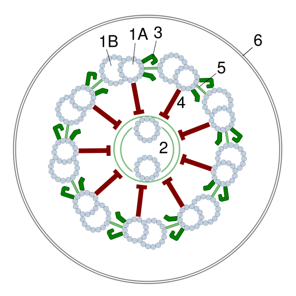
| Description | A diagrammatic cross-section of an axoneme as seen from the top of the undulipodia |
| Date | |
| Source | Own work based on: the descriptions and diagrams published in the textbooks of Cytology. |
| Author | Alexei Kouprianov |
| SVG development | This diagram was created with Sodipodi. This diagram uses embedded text that can be easily translated using a text editor. |
Un axonema consta de 9 pares de microtúbulos (A y B) exteriores que rodean un par central. A esta disposición se le conoce como 9x2+2
Licensing
I, the copyright holder of this work, hereby publish it under the following license:
This file is licensed under the Creative Commons Attribution-Share Alike 2.5 Generic license.
- You are free:
- to share – to copy, distribute and transmit the work
- to remix – to adapt the work
- Under the following conditions:
- attribution – You must give appropriate credit, provide a link to the license, and indicate if changes were made. You may do so in any reasonable manner, but not in any way that suggests the licensor endorses you or your use.
- share alike – If you remix, transform, or build upon the material, you must distribute your contributions under the same or compatible license as the original.
File history
Click on a date/time to view the file as it appeared at that time.
| Date/Time | Thumbnail | Dimensions | User | Comment | |
|---|---|---|---|---|---|
| current | 21:41, 15 September 2006 | | 656 × 656 (162 KB) | Alexei Kouprianov | {{Information |Description= A diagrammatic cross-section of an axoneme as seen from the top of the undulipodia |Source= Own work, based on the descriptions and diagrams published in the textbooks of Cytology. |Date= September, 16 2006 |Author= [[U |
File usage
No pages on the English Wikipedia use this file (pages on other projects are not listed).
Global file usage
The following other wikis use this file:
- Usage on bs.wikipedia.org
- Usage on ca.wikipedia.org
- Usage on de.wikipedia.org
- Usage on es.wikipedia.org
- Usage on fr.wikipedia.org
- Usage on gl.wikipedia.org
- Usage on hu.wikipedia.org
- Usage on ja.wikipedia.org
- Usage on nl.wikipedia.org
- Usage on pl.wikipedia.org
- Usage on pt.wikipedia.org
- Usage on ru.wikipedia.org
- Usage on uk.wikipedia.org